
Advanced Corneal Imaging and Keratoconus Monitoring
Modern keratoconus management relies heavily on advanced corneal imaging technologies.
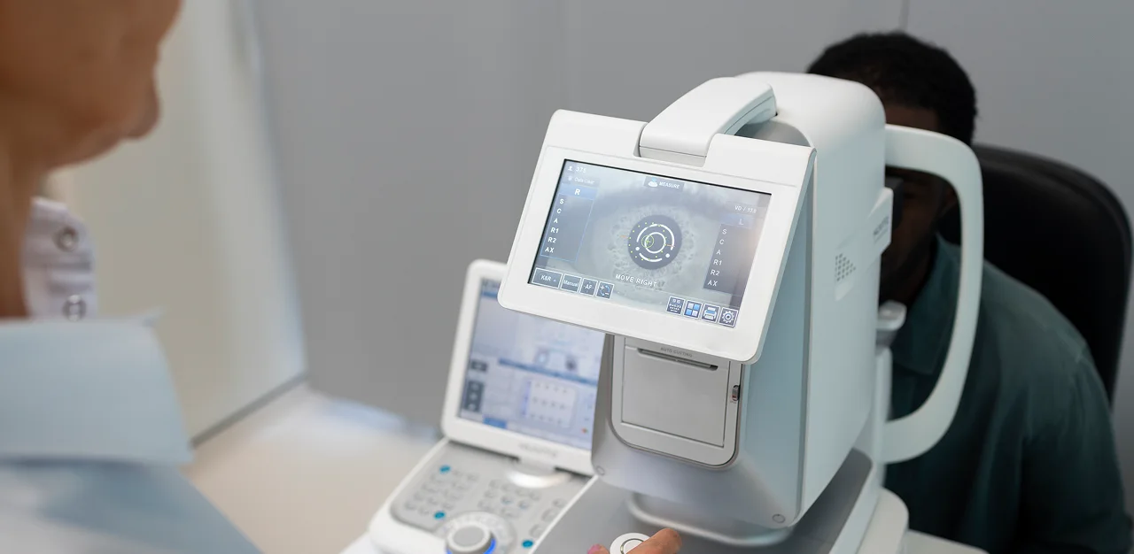
At Rose Optometry and the New Zealand Eye Research Centre (NZERC), multipleadvanced imaging systems are used to monitor corneal shape, detect progressionand guide treatment decisions.
Tomography is considered the gold standard for keratoconus monitoring andseverity assessment because it analyses both the front and back surfaces of thecornea together with corneal thickness distribution.
Technologies used include:
1-Pentacam AXL Wave
- Scheimpflug tomography
- Anterior and posterior elevation analysis
- Pachymetry progression mapping
- Belin/Ambrosio ectasia indices
- Axial length and wavefront analysis
2-Heidelberg Anterion
- Swept-source OCT tomography
- Epithelial and stromal assessment
- Advanced anterior segment imaging
- Early ectasia analysis
3-Medmont Meridia Pro
- Advanced corneal topography
- Orthokeratology analysis
- Dry eye and ocular surface evaluation
- Specialty contact lens fitting
4-Medmont Meridia Vantage
- Longitudinal corneal monitoring
- Orthokeratology analysis
- Scleral lens fitting support
- Specialty lens analysis
- Tear film assessment
5-ScanSys Tomographer
- Advanced corneal analysis
- Keratoconus assessment workflows
- Ectasia monitoring
- Specialty contact lens development support
Modern tomography systems allow clinicians to identify subtle posterior corneal changes before advanced visual symptoms develop.
This enables earlier intervention including:
- Corneal cross-linking
- Specialty contact lenses
- Long-term progression monitoring